Synovial sarcoma, left proximal calf
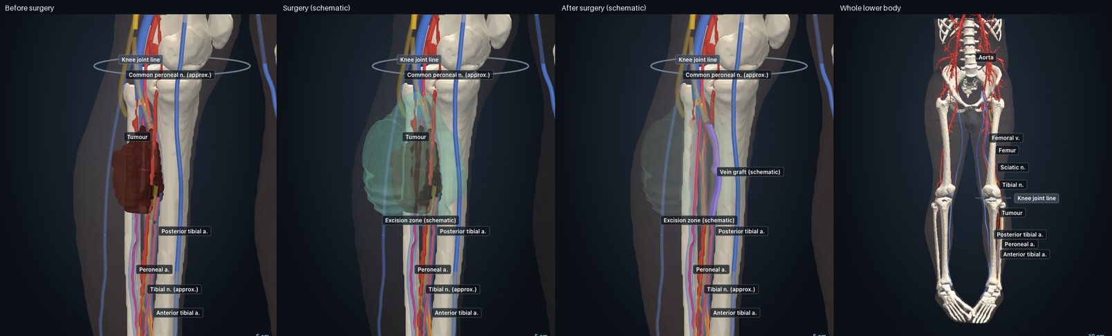
AI-generated 3D reconstruction (illustrative). Surgery and after views are schematic.
How the surgery was done
From the discharge summary (operation 9 Jun 2026) and the resection pathology (20 Jun 2026).
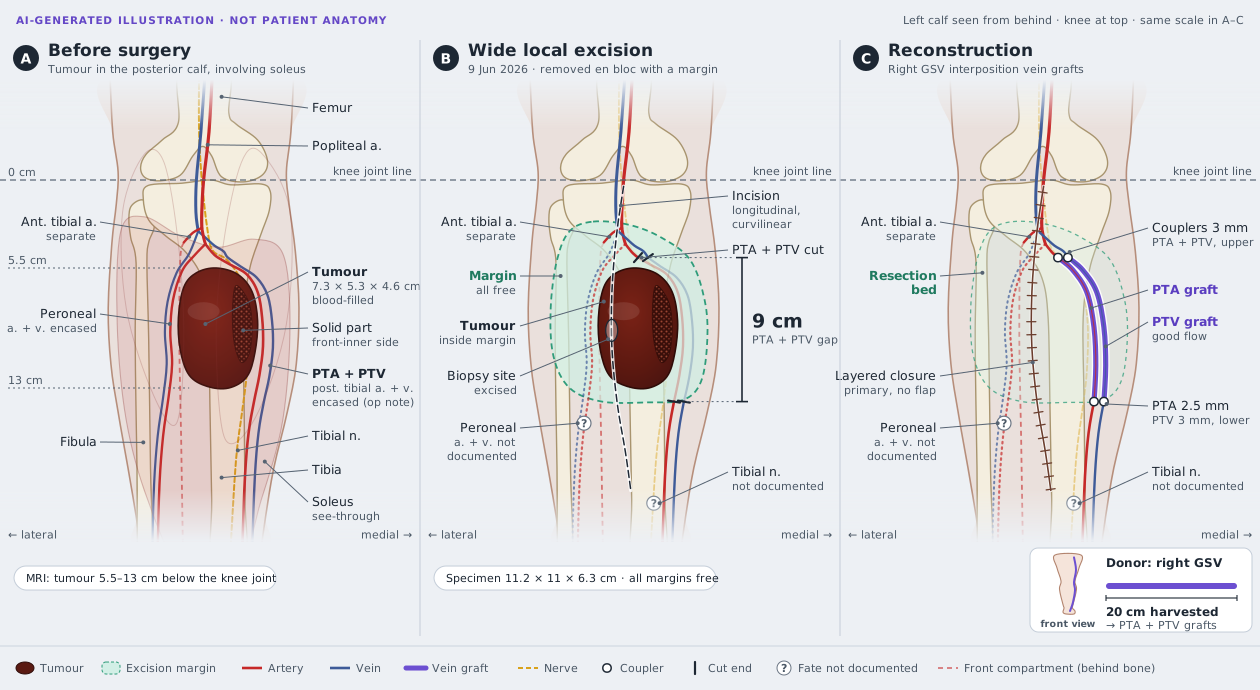
- 1Wide local excision through a long curved incision; the biopsy tract was removed with it. [Discharge summary]
- 2Posterior tibial and peroneal vessels were encased by the tumour; the anterior tibial vessels were separate. [Discharge summary]
- 3Posterior tibial artery and vein: a 9 cm gap between the stumps, bridged with interposition vein graft; 20 cm of the right great saphenous vein was harvested for it. Good flow afterwards. [Discharge summary]
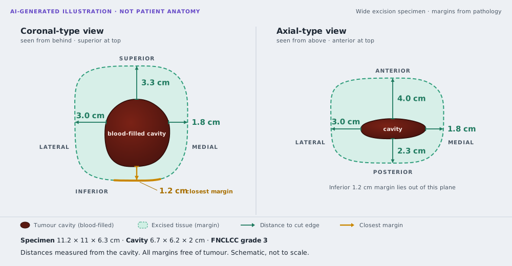
- 4Specimen 11.2 × 11 × 6.3 cm. All margins free of tumour; closest 1.2 cm (inferior). Grade 3, no lymphovascular invasion, no necrosis. [Histopathology] [Histopathology]
- ?Not documented: what happened to the peroneal vessels and the tibial nerve, and which muscles were removed.
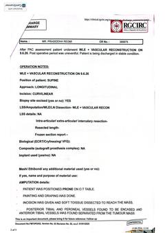 Discharge summary, page 2 of 5 (operation notes)2026-06-09
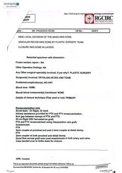 Discharge summary, page 3 of 5 (revascularisation)2026-06-09
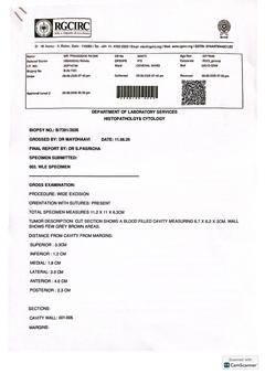 Histopathology: wide excision B/7391/2026, page 12026-06-09
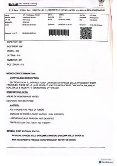 Histopathology: wide excision B/7391/2026, page 22026-06-09
Kathmandu MRI vs Delhi MRI
Kathmandu · 24 May
Sooriya Diagnostic, 1.5 T · AI calipers (report not in files)
Delhi · 27 May
RGCIRC, 3 T · radiologist [MRI left leg] · AI calipers 7.6 × 5.4–5.6 × 4.4–4.5 cm
Change in 3 days
AI observation Within measuring error: no measurable growth
What did change
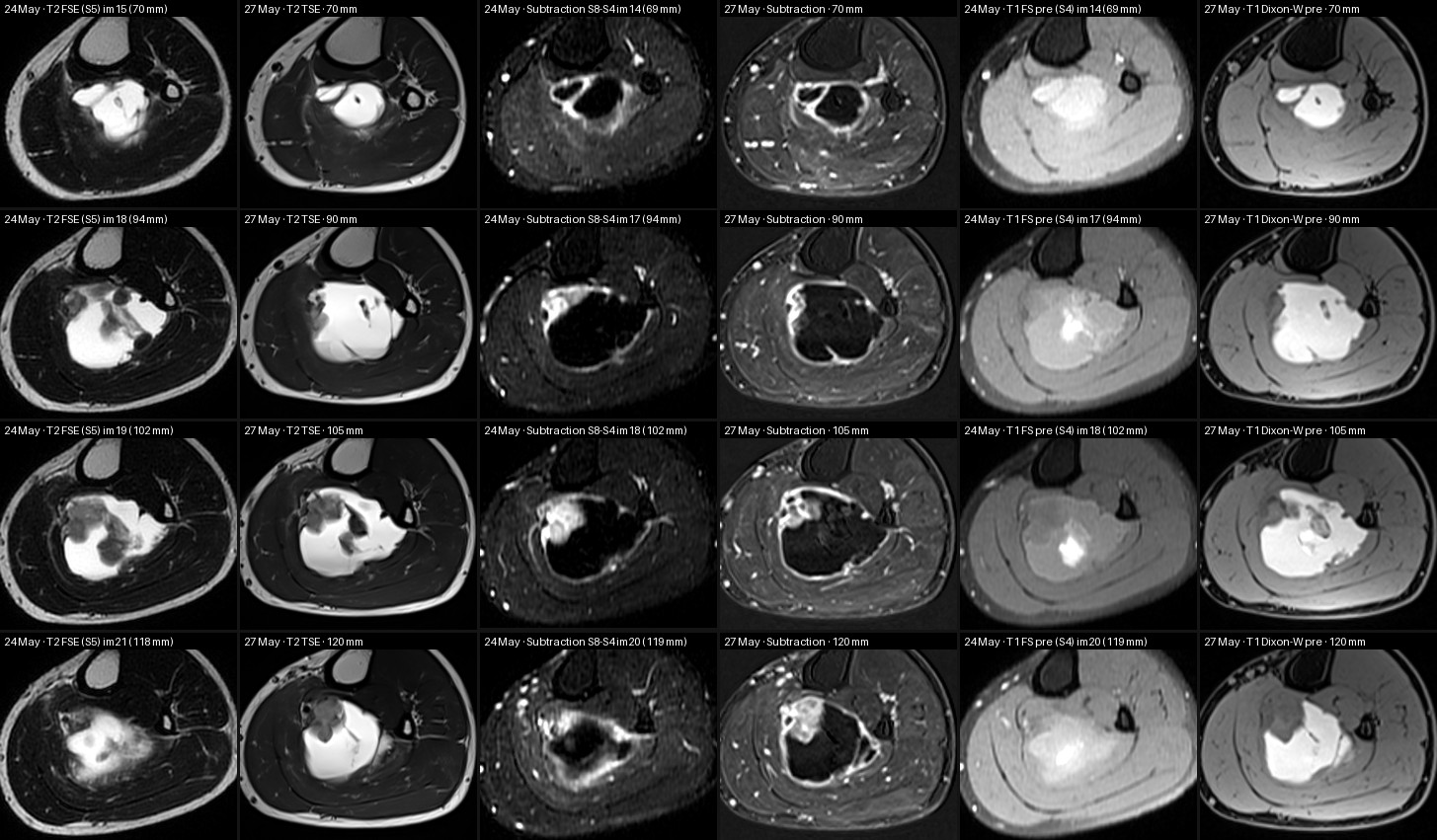
AI observation Dark compartments turned bright: most likely a recent bleed ageing rather than growth
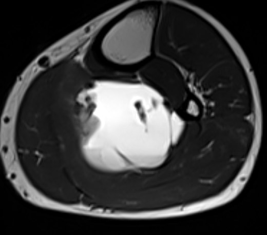
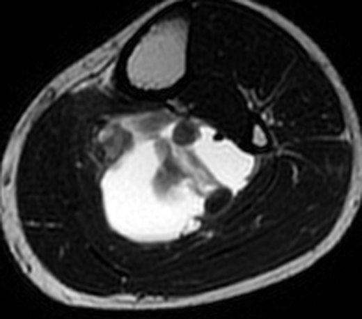
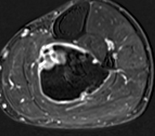
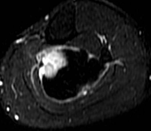
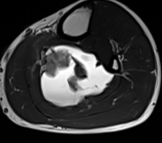
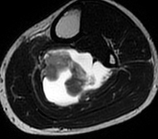
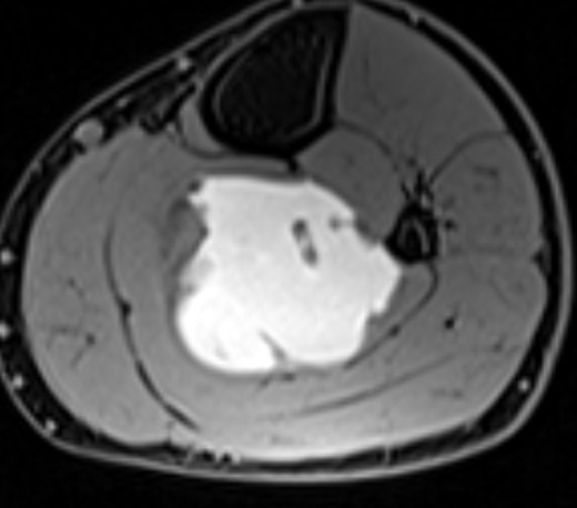
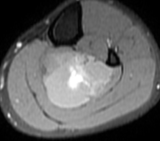
Kathmandu MRI images
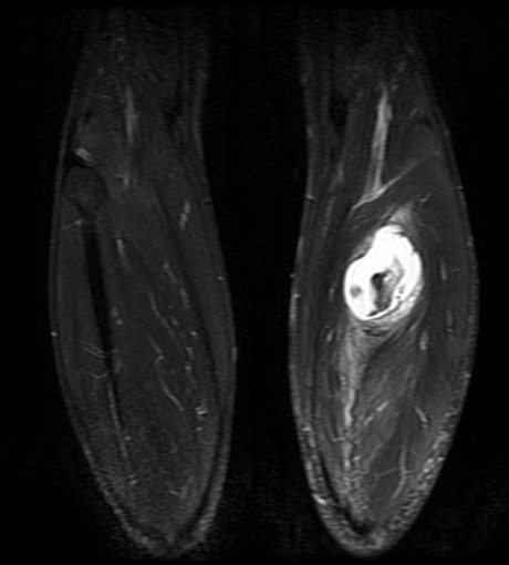
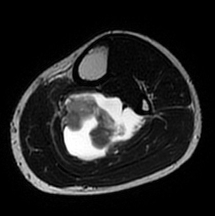
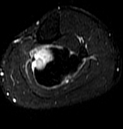
Scroll the original slices
Unannotated original images through the tumour. Drag on an image, use the slider, or click it and use the arrow keys or mouse wheel.
 RLAP
RLAP RLAP
RLAP RLHF
RLHF RLAP
RLAP RLAP
RLAP APHF
APHF RLHF
RLHF RLAP
RLAPWhere the tumour is
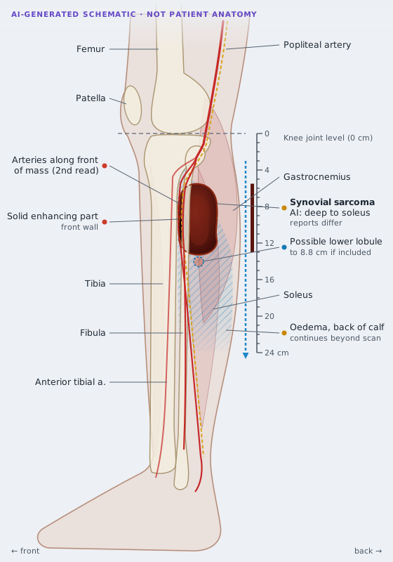
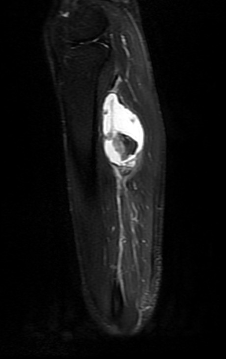
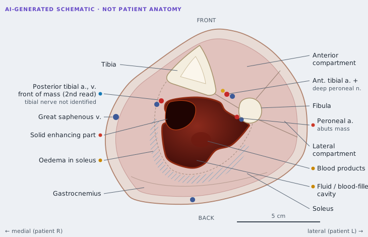
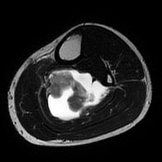
Each AI schematic sits next to the original image in the same orientation.
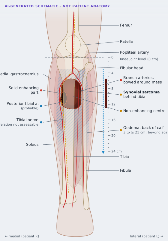
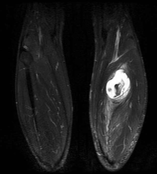
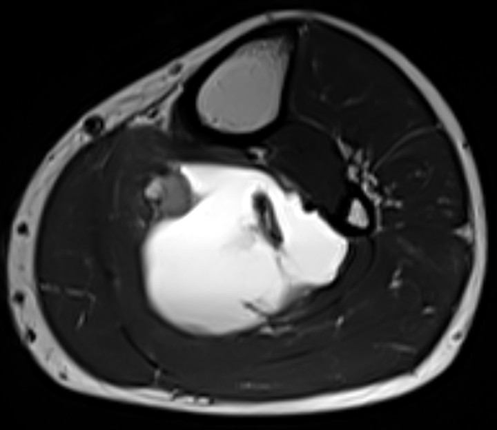
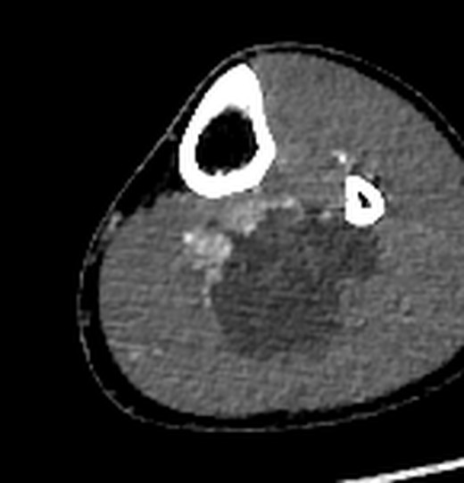
Has it spread?
Lung apices to upper abdomen. Non-contrast, low dose, breathing artefact.
Lung apices to upper abdomen. Non-contrast. No report in the files.
Diaphragm to proximal thighs. Non-contrast.
Renal arteries to toes, both legs. Arterial and late phases.
Pelvis to feet. Coronal STIR and Dixon, post-contrast subtraction.
Knee to ankle. Full multiparametric protocol.
Distal thigh to mid-calf (coronal STIR includes both legs).
Brain, head and neck, upper limbs. No PET-CT, bone scan or whole-body MRI.
| Region | Finding | Status |
|---|---|---|
| Lungs | June CT: no suspicious nodule [Low-dose CT chest]. June and September CT, two AI reads: no nodule of 5 mm or more, nothing new. | None reported Sep report pending |
| Lymph nodes | Abdomen: few sub-centimetre nodes, no lymphadenopathy [CT abdomen (whole)]. Groin ≤ 6.6 mm, pelvis ≤ 7.6 mm, behind knee ≤ 3.4 mm: AI measurements, not in any report. No node dissection at surgery. | None reported Groin, knee: AI only |
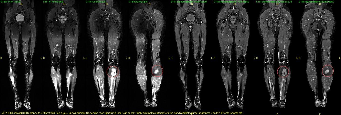
| Thighs and calves | Left leg: no satellite lesions [MRI left leg]. Right leg: no second lesion on AI review (no radiologist report). Swelling down the left calf to the ankle: AI observation (report: mild oedema around the mass). | None reported Right leg: AI only |
| Abdomen | Non-contrast CT: 3 mm left kidney stone only [CT abdomen (whole)]. | None reported |
| Bones | Small bone islands in both hips (harmless) [CT abdomen (whole)]. Tibia and fibula: normal signal and cortex [MRI left leg]; contact with the bone surface not excluded (AI). | None reported |
| Not scanned | Brain, arms; no PET-CT or whole-body MRI. | Not assessable |
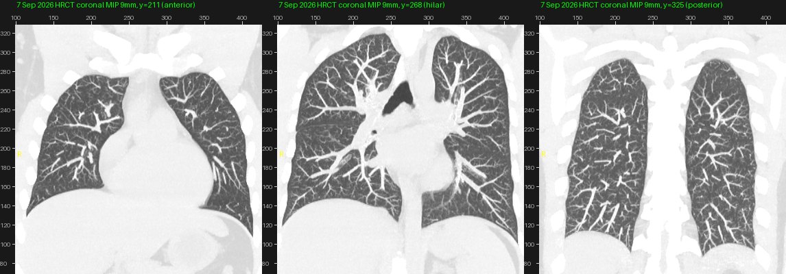
Pathology
| Biopsy · 29 May | Wide excision · 9 Jun | |
|---|---|---|
| Diagnosis | Synovial sarcoma | Residual spindle cell sarcoma: synovial sarcoma |
| Grade (FNCLCC) | At least 2 | 3 |
| Mitoses | ≈ 2–3 per 2 mm² | Brisk |
| Necrosis | Not seen | Not identified |
| IHC | TLE1+, TRPS1+, SS18+; CK, SMA, S100, MyoD1 negative; INI1 reduced | As biopsy |
| Margins | — | All free; closest 1.2 cm inferior (gross distances from the cavity) |
| Lymphovascular invasion | — | Not identified |
| Report | B/6802/2026, approved 2 Jun [Histopathology] | B/7391/2026, approved 20 Jun [Histopathology] [Histopathology] |
Molecular confirmation of the SS18::SSX fusion is not reported. Your phone shows the resection report as 3 pages; only 2 pages are in the files.
Documents and details
Scanned reports (select to enlarge). Detailed tables are folded below.
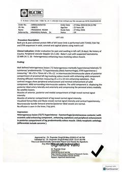 MRI left leg: report (RGCIRC)2026-05-27 · PDF p.1
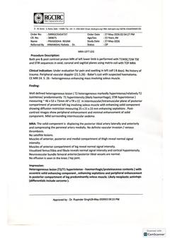 MRA left leg: report (RGCIRC)2026-05-27 · PDF p.2
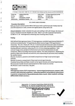 Bilateral lower-limb screening MRI: report2026-05-27 · PDF p.4
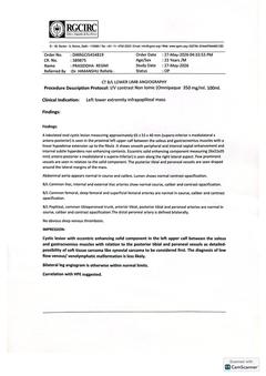 CT bilateral lower-limb angiography: report2026-05-27 · PDF p.3
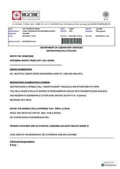 Histopathology: core biopsy B/6802/20262026-05-29 · PDF p.5
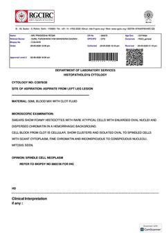 Cytology: aspirate C/2078/262026-05-29 · PDF p.6
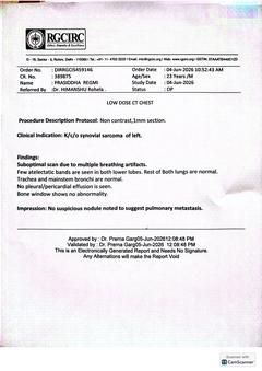 Low-dose CT chest: report2026-06-04 · PDF p.8
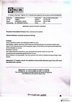 CT abdomen (whole), non-contrast: report2026-06-04 · PDF p.7
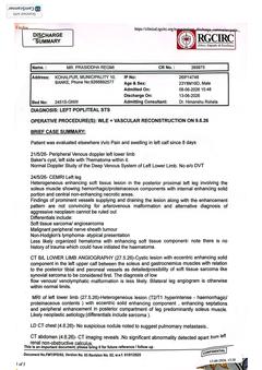 Discharge summary, page 1 of 52026-06-13 · PDF p.9Discharge summary, page 2 of 5 (operation notes)2026-06-09 · PDF p.11Discharge summary, page 3 of 5 (revascularisation)2026-06-09 · PDF p.10
 Discharge summary, page 4 of 5 (plan, medication)2026-06-13 · PDF p.12Histopathology: wide excision B/7391/2026, page 12026-06-09 · PDF p.13Histopathology: wide excision B/7391/2026, page 22026-06-09 · PDF p.14
Structure by structure (muscles, vessels, nerve, bone)
| Structure | Documented (treating team) | AI-assisted observation | Confidence | Surgical relevance |
|---|---|---|---|---|
| Level | Proximal left leg / upper calf; "left popliteal STS"; "infrapopliteal mass". [MRI left leg] [CT bilateral lower-limb angiography] [Discharge summary] | Top 5.5–5.6 cm and bottom 12.8–13.2 cm below the tibial plateau on both MRIs; 5.3 and 12.3 cm on CT. Upper pole just below the popliteal artery division, at the soleal arch. | High | Defines proximal margin relative to the popliteal bifurcation. |
| Compartment / plane | MRI: intermuscular/intramuscular, posterior compartment, involving soleus. CT: between soleus and gastrocnemius. [MRI left leg] [CT bilateral lower-limb angiography] | Deep to the soleus bulk, between soleus and the deep posterior compartment, against the posterior tibial and peroneal bundles (3 T T2, S19001). | Discrepancy Moderate | Determines resection planes; consistent with the operative finding of vessel encasement. |
| Soleus | Involved (MRI). | Stretched over the posterior surface with intramuscular oedema; origin within deep soleus not excluded. | Moderate | Extent of soleus resection not documented. |
| Gastrocnemius | Mass between soleus and gastrocnemius (CT). | Separated from the mass by the soleus on 3 T T2. | Discrepancy | Clarify with the radiologist. |
| Deep posterior compartment (TP, FDL, FHL) | Not described. | Displaced anteriorly; no intramuscular extension seen. | Moderate | |
| Posterior tibial vessels | Displaced laterally and anteriorly (MRI); draped around the lateral margins (CT); encased at surgery; 9 cm gap between PTA/PTV stumps bridged with interposition vein grafts. [Discharge summary] [Discharge summary] | Run along the anteromedial surface beside the enhancing solid component; patent. | Documented at surgery | Graft patency after surgery not documented. |
| Peroneal vessels | Compressed medially (MRI); draped around the lateral margins (CT); encased at surgery. [Discharge summary] | Run along the lateral margin near the fibula; patent. | Documented at surgery | Fate (ligated, preserved, reconstructed) not documented. |
| Anterior tibial vessels | Separated from the tumour (surgery). | Leave the popliteal artery above the mass and run in the anterior compartment. | Not involved | Separate from the tumour; preservation implied, not stated. |
| Tibial nerve | Not mentioned in any report. | Not separately identified on the available sequences. | Not assessable | Status and post-operative function to be confirmed. |
| Tibia | Normal signal and cortex (MRI). | Solid wall within a few millimetres of the posterior cortex at 9–11 cm; no marrow oedema or enhancement. | No invasion seen | Periosteal contact cannot be excluded on imaging. |
| Fibula | Linear hypodense extension up to the fibula (CT). | Approaches the posteromedial fibula; marrow normal. | Moderate | |
| Knee joint | No effusion in knee or hip (MRI). | Mass ends 5.5 cm below the joint; joint not involved. | Not involved | |
| Skin and subcutis | Not described. | Not involved. | Not involved | |
| Peritumoral oedema | "Mild surrounding intermuscular oedema" (MRI). | Posterior-compartment oedema from about 3 cm below the joint to the ankle on the 27 May whole-leg STIR; anterior and lateral compartments spared. | Extent Cell content | Tumour cells within oedema cannot be assessed on MRI. |
Tibial nerve (AI review): not separately identified at the tumour level on CT or MRI; its course in the 3D model is approximated. Confidence low.
Measurements by study
All tumour measurements across studies, with the method behind each number. Craniocaudal (CC), transverse (TR, medial-lateral) and anteroposterior (AP).
| Study | Source | What was measured | CC (cm) | TR / ML (cm) | AP (cm) | Solid component |
|---|---|---|---|---|---|---|
| Pre-operative imaging (all before the 29 May biopsy and aspiration) | ||||||
| MRI 24 May · outside 1.5 T | AI observation | Outer enhancing wall, calipers on subtraction (S10 im10, S9 im14, S8 im17) | 7.5–7.8 (8.8 incl. possible inferior lobule) | 5.1–5.2 | 4.2–4.5 | ≈2–3 cm, anteromedial |
| MRI 24 May · outside 1.5 T | AI observation | Fluid component, semi-automatic STIR outline | 7.3 | 5.2 | 4.4 | — |
| MRI 27 May · RGCIRC 3 T | Documented [MRI left leg] | Radiologist (whole lesion) | 7.3 | 5.3 | 4.6 | 25 × 21 × 23 mm (axes not stated), diffusion restriction |
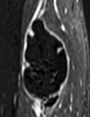
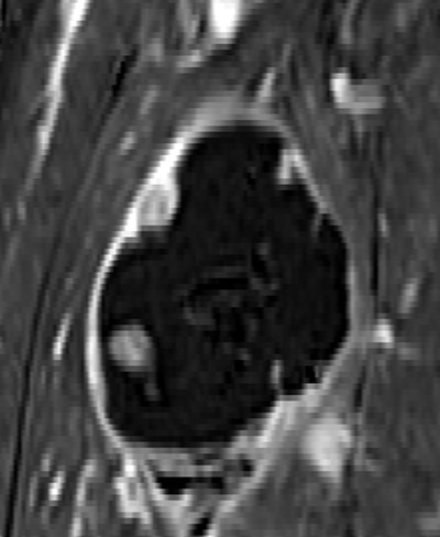
| MRI 27 May · RGCIRC 3 T | AI observation | Outer wall, calipers on 3D Dixon subtraction (S1014001) | 7.6 (sagittal) | 5.4–5.6 | 4.4–4.5 | 2.2 (AP) × 2.1 (TR) × 3.2 (CC), ADC median 1.12 ×10⁻³ mm²/s |
| MRI 27 May · RGCIRC 3 T | AI observation | Fluid component, semi-automatic STIR outline (≈57 mL) | 7.5 | 5.2 | 4.2 | — |
| CT angiography 27 May | Documented [CT bilateral lower-limb angiography] | Radiologist | 6.5 | 5.3 | 4.0 | 2.6 (AP) × 2.1 (ML) × 3.5 (SI) |
| Pathology (after aspiration of 50 mL on 29 May) | ||||||
| Resection 9 Jun | Documented [Histopathology] | Blood-filled cavity 6.7 × 6.2 × 2 cm (axes not stated); specimen 11.2 × 11 × 6.3 cm | — | — | — | Wall shows few grey-brown areas |
Values differ mainly by what is measured (outer wall, fluid cavity or whole lesion), by plane and by modality. The 29 May aspiration yielded 50 mL of blood with clot; the resected cavity's smallest dimension (2.0 cm, axis not stated) is below every pre-operative measurement, consistent with decompression and specimen shrinkage. AJCC 8th edition size category from the documented maximum diameters (7.3 cm MRI, 6.5 cm CT): T2 (more than 5 cm, up to 10 cm); the AI calipers (7.5–7.6 cm) give the same category.
MRI 24 May vs MRI 27 May AI observation
| Feature | 24 May (GE 1.5 T, outside) | 27 May (Siemens 3 T, RGCIRC) | Interpretation |
|---|---|---|---|
| Size (outer wall) | 7.5–7.8 × 5.1–5.2 × 4.2–4.5 cm | 7.6 × 5.4–5.6 × 4.4–4.5 cm | No measurable change (differences ≤ 5 mm, within method error of ±5 mm) |
| Position | Upper pole 5.5 cm, lower pole 12.8 cm below joint | 5.6 and 13.2 cm | Unchanged within error |
| T2 internal signal | Several T2-dark compartments with fluid levels | Most compartments T2-bright | Evolution of subacute haemorrhage (intracellular to extracellular methaemoglobin) |
| T1 signal before contrast | Focal T1-bright (fat-suppressed) area | T1 hyperintensity through most of the cavity | Methaemoglobin; partly sequence-related (2D spin echo vs 3D Dixon gradient echo) |
| Solid component | Anteromedial enhancing nodule, about 2–3 cm | Anteromedial, 2.1 × 2.2 × 3.2 cm, restricted diffusion | Same position; size comparable within limits |
| Enhancement | Thick irregular rim, septa, nodular component | Rim, septa, nodular component (minimal solid enhancement per report) | Similar |
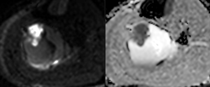
| Diffusion (ADC) | Solid part 1.7–2.0 ×10⁻³ mm²/s (b-values not stored) | Solid part ≈1.1 ×10⁻³; cavity ≈2.7 ×10⁻³ | Not comparable: different b-values, field strength and resolution |
| Oedema | Posterior compartment to the field edge (~21 cm below joint) | Posterior compartment to the ankle | Similar where both studies overlap; full extent only on 27 May |
| Vessels | Arteries displaced, patent; tumour blush (TRICKS) | PTA/peroneal displaced, patent; tumour blush (TWIST, CTA) | Similar |
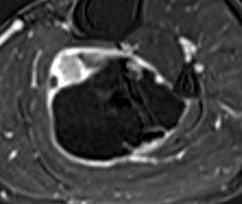
Progression and aggressiveness
Progression. AI observation No measurable growth between 24 and 27 May 2026. Three days is too short to estimate growth kinetics, and the scans differ in scanner, field strength and protocol. The visible change is in the blood products inside the tumour, consistent with a bleed that began days before 24 May and fits the acute presentation. No imaging of the leg after the 9 Jun resection is in the files, so local recurrence cannot be assessed. The 7 Sep 2026 HRCT shows no new lung nodule on AI review; it does not cover the leg.
Aggressiveness. Documented high-risk features: synovial sarcoma, FNCLCC grade 3 with brisk mitoses [Histopathology], deep location, size above 5 cm, encasement of the posterior tibial and peroneal vessels [Discharge summary]. Imaging features AI observation: haemorrhage, solid component with restricted diffusion, peritumoral oedema to the ankle, tumour hypervascularity. Favourable documented features: all margins free of tumour (closest 1.2 cm), no lymphovascular invasion, no necrosis, no suspicious lung nodule on the June staging CT. Validated tools such as the Sarculator nomogram estimate prognosis from these variables; that estimate belongs to the treating team.
Vessels, one by one
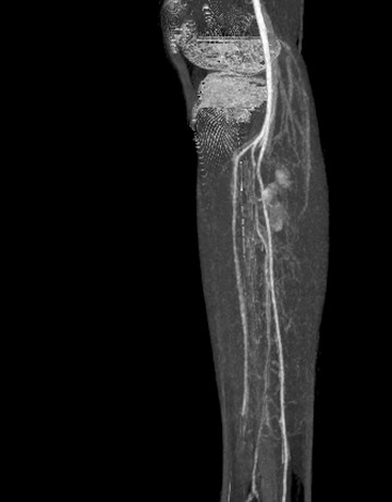
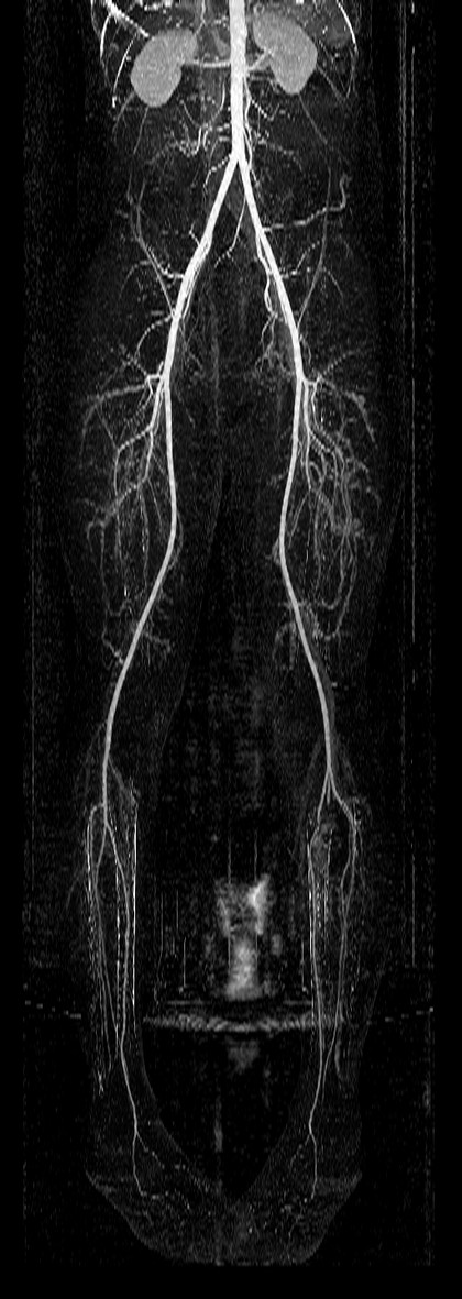
Vessel-by-vessel review combining the CT angiogram, both MRAs and the operative record.
| Vessel | Study | Documented | AI-assisted observation | Uncertainty / follow-up |
|---|---|---|---|---|
| Aorta, iliac, common/superficial/deep femoral (both) | CTA 27 May | Normal course, calibre, opacification [CT bilateral lower-limb angiography] | Patent on subtraction MIP | — |
| Popliteal artery (L) | CTA 27 May | Normal | Patent; divides just below the knee joint, above the tumour | — |
| Anterior tibial artery (L) | CTA · surgery | Normal (CT); vessels separated from tumour (surgery) | Leaves above the mass, runs in the anterior compartment; patent to the ankle | Separate from tumour; preservation implied, not stated |
| Tibioperoneal trunk (L) | CTA 27 May | Normal | At the upper pole of the mass | Relation to the resection not documented |
| Posterior tibial artery (L) | MRI · CTA · surgery | Displaced laterally and anteriorly by the solid component (MRI); draped around the lateral margins of the mass (CT); encased (surgery); 9 cm gap between PTA/PTV stumps bridged with an interposition vein graft [Discharge summary] [Discharge summary] | Along the anteromedial surface beside the enhancing solid component; patent, no cut-off | Graft: right GSV interposition, 3 mm coupler proximal, 2.5 mm distal; post-operative patency not documented |
| Posterior tibial vein(s) (L) | Surgery | Encased; reconstructed with interposition vein graft, 3 mm couplers both ends [Discharge summary] | Not separately assessed | Post-operative patency not documented |
| Peroneal artery (L) | MRI · CTA · surgery | Compressed medially (MRI); draped around the lateral margins (CT); encased (surgery) | Along the lateral margin near the fibula; patent | Fate not documented; not listed as reconstructed |
| Peroneal veins (L) | Surgery | Encased | Not assessed | Fate not documented |
| Deep veins (L) | Doppler 21 May · CTA · MRA | Doppler: no evidence of DVT; CTA: no obvious deep venous thrombosis; MRA: no definite vascular invasion or venous thrombosis [Discharge summary] [CT bilateral lower-limb angiography] [MRA left leg] | — | — |
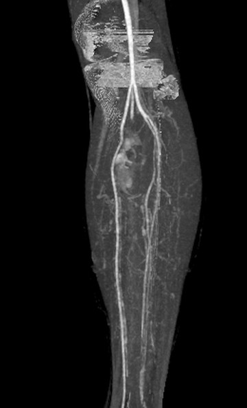
| Distal run-off (L) | CTA 27 May | "The distal peroneal artery is defined bilaterally"; angiogram otherwise within normal limits | Anterior tibial, posterior tibial and peroneal arteries opacify down the calf on the subtraction MIP (pre-operative) | Post-operative run-off not imaged |
| Tumour vascularity | MRI 24 May · CTA | Prominent supplying and draining vessels (24 May summary); few prominent vessels at the solid component (CT) | Early blush centred on the anteromedial solid component (TRICKS, TWIST, arterial CT) | — |
Discrepancy Pre-operative imaging described displacement without definite invasion; surgery found the posterior tibial and peroneal vessels encased. The same 27 May MRI reports also state that the neurovascular bundle vessels are normal, which conflicts with their own displacement description. The MRI wording (posterior tibial artery displaced laterally, peroneal compressed medially) is unusual because the posterior tibial artery normally lies medial to the peroneal. On the images the posterior tibial artery runs along the anteromedial surface and the peroneal along the lateral surface. Whether the vessels can be preserved or reconstructed is a decision for the vascular and surgical teams.
Nerves, muscle, bone, joint
Each structure is classed as documented, suspicious, not identified or not assessable. Not identified does not mean confirmed absent.
Muscles and compartments
- Involved Posterior compartment: soleus involved [MRI left leg]; AI review: soleus stretched over the mass, deep compartment displaced.
- Not identified Anterior compartment of the leg normal Documented [MRI left leg]; lateral compartment normal on AI review.
- Not identified Thigh muscles (anterior, posterior, medial) normal Documented [MRI left leg]. AI review of both thighs on CT angiography and coronal MRI found no mass or asymmetric muscle.
- Oedema Posterior-compartment oedema to the ankle (27 May). Oedema is not tumour, but its cell content cannot be assessed on MRI.
Nerves
- Not assessable Tibial nerve: not mentioned in imaging, operative or pathology reports; not separately resolved on the available sequences. It runs with the posterior tibial vessels, which were encased.
- AI observation Common peroneal nerve: seen in the popliteal fossa on AI review; not separately resolved at the fibular neck. Not documented in reports.
Bones and joints
- Not identified Femur, tibia and fibula: normal signal and cortex Documented [MRI left leg]; no marrow oedema or enhancement next to the mass on AI review.
- Contact Solid wall within a few millimetres of the posterior tibial cortex; periosteal contact cannot be excluded.
- Not identified No knee or hip effusion Documented. Tiny sclerotic foci in both femoral heads, read as bone islands Documented [CT abdomen (whole)].
Skin and superficial tissue
- Not identified No skin or subcutaneous involvement on imaging.
- Curvilinear longitudinal incision, biopsy site excised, primary closure Documented [Discharge summary]. Wound status after discharge is not documented.
Full staging table
| Region | Imaged by | Documented | AI-assisted observation | Status |
|---|---|---|---|---|
| Chest | ||||
| Lungs | LDCT 4 Jun 2026 (pre-op) · HRCT 7 Sep 2026 (post-op) | LDCT: "Suboptimal scan due to multiple breathing artifacts." "Few atelectatic bands are seen in both lower lobes. Rest of Both lungs are normal." Impression: "No suspicious nodule noted to suggest pulmonary metastasis." [Low-dose CT chest] HRCT: no report in the files. | Two independent reads (lung segmentation, blob detection with synthetic-nodule sensitivity testing, 9 mm MIP review, 1 mm confirmation): no nodule ≥ 5 mm on either date; no new or enlarging opacity from June to September. Detection of 3–4 mm nodules: June lower lobes poor (motion), September 6–7 of 8. | Not identified < 5 mm not excluded |
| Airways, pleura, pericardium | LDCT · HRCT | "Trachea and mainstem bronchi are normal. No pleural/pericardial effusion is seen." [Low-dose CT chest] | Airways patent; no effusion, pneumothorax or pericardial effusion on either date. No discrete band atelectasis found by AI review; mild dependent density in June from a shallow breath, resolved in September. | Not identified |
| Mediastinal, hilar, axillary, supraclavicular nodes | LDCT · HRCT (non-contrast) | Not mentioned in the LDCT report. | No node ≥ 10 mm short axis. Non-contrast limits separation of small nodes from vessels. Anterior mediastinal soft tissue fits normal thymus for age. | Not identified Uncertain |
| Abdomen and pelvis | ||||
| Liver, spleen, pancreas, adrenals | CT abdomen 4 Jun 2026 (non-contrast) · CTA 27 May (upper abdomen) | "Liver is normal on plain scan." No dilated intrahepatic biliary radicles. "Gall bladder, spleen, pancreas, both adrenals and both kidneys are normal on plain scan." [CT abdomen (whole)] | Liver homogeneous (~61 HU); spleen ~10 cm; adrenals thin. Small lesions cannot be excluded without contrast. | Not identified Non-contrast |
| Kidneys, ureters, bladder | CT abdomen 4 Jun | "About 3 mm calculus seen at the interpolar region of left kidney. No hydroureteronephrosis." [CT abdomen (whole)] | 2 mm hyperdense focus, mid left kidney (~370 HU), no hydronephrosis; kidneys 9.4 cm (R), 10.3 cm (L). | Not identified |
| Bowel, peritoneum, prostate | CT abdomen 4 Jun | "Urinary bladder is normal. Prostate is normal." No ascites. | Unremarkable. | Not identified |
| Para-aortic, iliac, mesenteric nodes | CT abdomen 4 Jun · CTA 27 May | "Few subcm discrete mesenteric and retroperitoneal lymph nodes are seen. No retroperitoneal lymphadenopathy/ascites is seen." | None enlarged: para-aortic ≤ 6.7 mm, external iliac 5.1 (R) / 7.6 mm (L), mesenteric ≤ 5 mm short axis. Size alone cannot exclude micrometastasis. | Not identified |
| Lower limbs (patient's focus: thighs and calves) | ||||
| Inguinal nodes | CTA 27 May · CT abdomen 4 Jun · MRI 27 May | Not mentioned in any report. | Largest 6.4 mm (R) and 6.6 mm (L) short axis; symmetric; normal fatty hila where visible. | Not identified |
| Lymph node sampling | Operation 9 Jun · resection pathology | No node dissection (WLE + vascular reconstruction only) [Discharge summary]; the pathology report does not mention lymph nodes [Histopathology]. | — | Pathological N not assessed |
| Popliteal nodes | CTA 27 May · MRI 24/27 May | Not mentioned. | None above 3.4 mm on either side. | Not identified |
| Thighs, both | CTA 27 May (whole length) · MRI 27 May coronal STIR/Dixon/subtraction (pelvis to feet) | Muscles of the anterior, posterior and medial compartments of the thigh normal. [MRI left leg] | No mass, no asymmetric muscle, no abnormal enhancement, femora normal on CT and MRI (two independent reads). The brighter left buttock and upper thigh on MRI is a receiver-coil effect: same on T1 and fat images, symmetric muscle density on CT (54–57 HU). | Not identified < 5–7 mm not excluded |
| Right calf | CTA 27 May · MRI 27 May coronal | Not separately reported; the bilateral screening report repeats the left-leg text. | No mass; bones normal. Symmetric STIR bands in both upper calves, not mass-like and absent on T1 and CT: likely technical or non-specific. | Not identified |
| Left calf outside the primary | MRI 24 and 27 May · CTA 27 May | "No satellite lesions." "Mild surrounding intermuscular oedema." [MRI left leg] | No separate nodule or skip lesion. Feathery high STIR signal fills the deep posterior compartment and soleus, continuous from the mass to at least 11 cm below its lower pole, with lower CT muscle density on the left (48–49 vs 54–55 HU): oedema most likely. Tumour spread along fascia cannot be excluded on imaging. | Oedema extent Microscopic spread |
| Bones of pelvis, femora, tibiae, spine (L) | CT abdomen 4 Jun · CTA 27 May | LDCT: "Bone window shows no abnormality." CT abdomen: "Bone window is unremarkable. Few tiny sclerotic foci noted in bilateral femoral heads- bony islands." [Low-dose CT chest] [CT abdomen (whole)] | Three 3–4 mm bone islands in the femoral heads, identical on both CTs; no lytic or blastic lesion. Thin transverse lines in both distal tibiae: growth-arrest lines. | Not identified |
| Never imaged | ||||
| Brain, head and neck, upper limbs; whole-body | None | Not requested in the files. | No PET-CT, bone scan or whole-body MRI. | Not assessable |
Surgery record (full)
Taken from the discharge summary, the only operative record in the files. It describes the operation already performed; it is not a plan.
- Date
- 9 Jun 2026 (admitted 8 Jun, discharged 13 Jun) [Discharge summary]
- Procedure
- Wide local excision of the left calf mass + vascular reconstruction (diagnosis recorded as "left popliteal STS")
- Teams
- Orthopaedic team: operating surgeon not named on the scanned pages (admitting consultant Dr Himanshu Rohela). Plastic surgery: "Surgeon(s) involved: DR RAJAN ARORA AND TEAM"; revascularisation scrub team Dr Rajan, Dr Amit; "Case handed over to Ortho team for closure."
- Approach
- Longitudinal, curvilinear incision; biopsy site excised
- Position
- "Supine" in the template field, "prone on OT table" in the narrative Discrepancy
- Findings
- Posterior tibial and peroneal vessels encased; anterior tibial vessels separate from the tumour [Discharge summary]. "Other operative findings: NA" [Discharge summary]
- Reconstruction
- 9 cm gap between PTA and PTV stumps; 20 cm right GSV harvested; PTA and PTV revascularised with interposition vein graft; PTA couplers 3 mm proximal and 2.5 mm distal; PTV 3 mm both ends; "Good flow across graft seen post anastomosis in both artery and veins." [Discharge summary]
- Closure
- "Closure was done in layers"; primary (no flap); blood loss 150 mL; no transfusion; "Problems/complication(s): NO ANY" [Discharge summary]
- Frozen section
- "NA"; HPE "Awaited" at discharge
- Node dissection
- None: "LSS/Amputation/WLE/LN Dissection- WLE + VASCULAR RECON" [Discharge summary]
- Template
- The operative narrative sits under the template heading "AMPUTATION details:"; no amputation was performed.
Not documented
- Fate of the encased peroneal artery and veins.
- Tibial nerve: dissected, preserved or sacrificed.
- Which muscles were resected (soleus, deep compartment) and the planes used.
- Specimen orientation details beyond "sutures present".
- Post-operative wound, graft surveillance, distal pulses, neurological status after 13 Jun 2026.
AI observation Post-operative anatomy cannot be inferred from the images here: every image of the leg predates surgery.
Discharge advice as printed: Tab Ceftum 500 mg twice daily × 7 days; Tab Ultracet twice daily × 7 days; Tab Pan 40 mg once daily × 7 days; Xyteen Pro protein powder 2 tsf thrice daily × 7 days; Tab Nerwita GN 400 mg HS × 5 days [Discharge summary]. Proposed plan: "To be decided after HPE report"; next visit 19 Jun 2026. "Patient is being discharged in stable condition" [Discharge summary].
Pathology (full)
Biopsy, cytology and resection reports side by side.
| Biopsy B/6802/2026 [Histopathology] | Cytology C/2078/26 [Cytology] | Resection B/7391/2026 [Histopathology] [Histopathology] | |
|---|---|---|---|
| Specimen | Core biopsy, multiple cores 0.3–1.3 cm | Aspirate 50 mL blood with clot (cell block) | Wide excision, oriented with sutures; 11.2 × 11 × 6.3 cm |
| Morphology | Spindle cells in short fascicles and storiform pattern; stellate hyperchromatic nuclei | Foamy histiocytes, rare atypical cells; cellular cell block of oval to spindled cells | Ill-defined spindle-cell tumour in short fascicles; coarse chromatin, prominent nucleoli |
| Mitoses | ≈2–3 / 2 mm² | Mitosis seen | Brisk |
| Necrosis | Not seen | — | Not identified |
| IHC | TLE1+, TRPS1+, SS18+; CK−, SMA−, S100−, MyoD1−; INI1 downregulated | Refers to biopsy | Refers to biopsy |
| Diagnosis / grade | Synovial sarcoma, at least FNCLCC grade 2 | Spindle-cell neoplasm | "Residual spindle cell sarcoma: synovial sarcoma (FNCLCC grade 3)" |
| Tumour | — | — | Blood-filled cavity 6.7 × 6.2 × 2 cm; wall shows few grey-brown areas; areas of haemorrhage |
| Margins | — | — | "All margins are free of tumor." Gross distance from cavity to margins: superior 3.3, inferior 1.2, medial 1.8, lateral 3.0, anterior 4.0, posterior 2.3 cm. Microscopic: closest margin 1.2 cm (inferior) |
| LVI | — | — | Not identified |
| Pre-resection therapy | — | — | None |
| Signatories | Case seen by Dr Meenakshi, Dr S. Pasricha, Dr A. Sharma; referred by Sunil Puri/Bharat/KM Mahendra/Gaurav; approved 2 Jun 2026 | Approved 2 Jun 2026 | Grossed by Dr Maydhaavi (11 Jun); final report Dr S. Pasricha; approved 20 Jun 2026 |
Grade. The biopsy grade of "at least 2" was raised to 3 on the resection specimen; core biopsies often under-grade. The component scores (differentiation, mitotic count per 10 HPF, necrosis) are not itemised in the report.
Molecular. Not documented The report lists SS18 expression; this is most likely the SS18-SSX fusion-specific antibody (interpretation; clone not stated), which strongly supports the diagnosis. Neither the clone nor any FISH, RT-PCR or sequencing confirmation of the SS18::SSX fusion is reported. The histological subtype (monophasic, biphasic, poorly differentiated) is not stated.
Margins. The gross distances are reported from the cavity; the microscopic line gives the closest margin as 1.2 cm (inferior) without stating its reference point. The report says "all margins are free of tumor" and does not use the term R0. The tissue at each margin (fascia, periosteum, vessel adventitia) is not described. No lymph nodes were submitted.
Necrosis. Imaging described non-enhancing, "necrotic" areas; pathology found a blood-filled cavity and no necrosis. The non-enhancing contents were haemorrhage, which matches the MRI signal (section 5).
Timeline
All dates 2026. Imaging events reference the report; the image review itself is in sections 4 to 8.
- Symptom onsetPain and swelling of the left calf for about 8 days before evaluation; no trauma. Exact date not documented.
- Venous Doppler, left leg (outside)Baker's cyst on the left with a possible haematoma; deep veins normal, no DVT. Report not in files.
- Contrast MRI, left leg (Sooriya Diagnostic, Kathmandu, 1.5 T)Heterogeneous enhancing lesion of the posterior proximal left leg involving the soleus, with haemorrhagic/proteinaceous components, internal enhancing solid portion and central non-enhancing areas; prominent supplying and draining vessels, not convincing for arteriovenous malformation. Differentials: soft-tissue sarcoma/angiosarcoma, MPNST, atypical non-Hodgkin lymphoma; less likely organised haematoma. DICOM reviewed here.
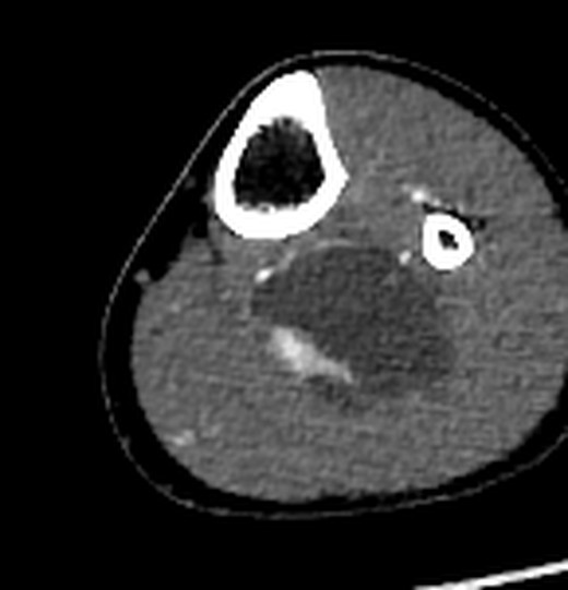
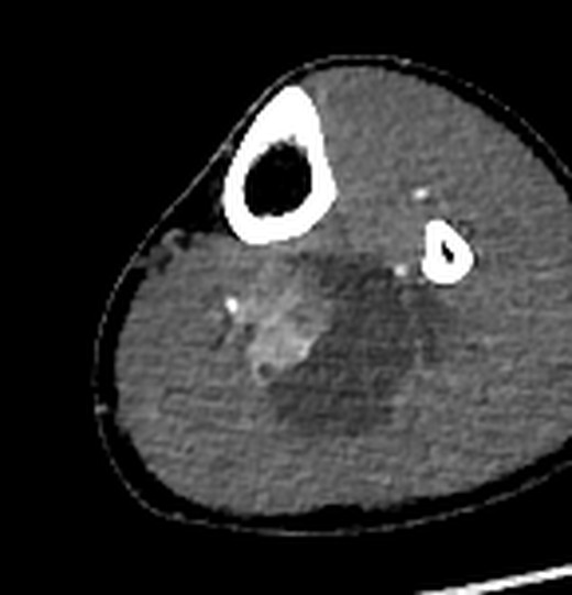
- CT angiography both legs (RGCIRC)Lobulated cystic lesion 65 × 53 × 40 mm (SI × ML × AP) between soleus and gastrocnemius; eccentric enhancing solid component 26 × 21 × 35 mm (AP × ML × SI); posterior tibial and peroneal vessels draped around the lateral margins of the mass. Synovial sarcoma to be considered first; low-flow venous/venolymphatic malformation less likely; angiogram otherwise normal.
- MRI left leg, MRA, bilateral screening (RGCIRC, 3 T)46 × 53 × 73 mm (AP × TR × CC), posterior compartment involving soleus; solid component 25 × 21 × 23 mm with diffusion restriction; posterior tibial artery displaced laterally and anteriorly, peroneal compressed medially; MRA: no definite vascular invasion or venous thrombosis; no satellite lesions. Impression: likely neoplastic (differentials include sarcoma).
- Core biopsy and aspirationBiopsy B/6802/2026; aspirate C/2078/26 yielded 50 mL of blood with clot.
- Biopsy reportedSynovial sarcoma, at least FNCLCC grade 2 (TLE1, TRPS1, SS18 positive).
- Staging CT chest and abdomenChest: no suspicious nodule to suggest pulmonary metastasis (suboptimal, breathing artefacts; few lower-lobe atelectatic bands). Abdomen (non-contrast): no significant abnormality apart from a 3 mm non-obstructive left renal calculus; few sub-centimetre nodes.
- AdmittedIP 26IP14748.
- Wide local excision + vascular reconstructionPosterior tibial and peroneal vessels encased; PTA and PTV reconstructed with right GSV interposition graft; biopsy site excised.
- DischargedPost-operative period uneventful; discharged in stable condition. Plan to be decided after the HPE report; next visit 19 Jun 2026.
- Resection histopathology approvedResidual spindle cell sarcoma: synovial sarcoma (FNCLCC grade 3); all margins free of tumour, closest 1.2 cm (inferior); no LVI; no pre-resection therapy.
- Adjuvant treatmentNot documented in the files. The request mentions "IA" cycles 1 and 2 (most likely ifosfamide with doxorubicin; intra-arterial chemotherapy is the other reading).
- HRCT chest (RGCIRC)Non-contrast HRCT, 3 months after surgery; no report in the files. AI review: no pulmonary nodule of 5 mm or more, no change since 4 Jun 2026.
Records inventory
Every conclusion in this dossier traces to one of these sources. Exam dates and report-approval dates are listed separately where they differ.
- Patient
- PRASIDDHA REGMI · male
- Hospital ID
- CR No. 389875 (RGCIRC, Delhi)
- Age / DOB
- DOB 25 Sep 2002 in every RGCIRC DICOM header; 23 y in all RGCIRC reports. The 24 May MRI header (Sooriya Diagnostic, Kathmandu) records 25 y (section 13).
- Treating team
- Admitting and referring consultant Dr Himanshu Rohela. Plastic surgery (Dr Rajan Arora and team; scrub team Dr Rajan, Dr Amit) performed the vascular reconstruction and handed the case to the orthopaedic team for closure. The orthopaedic operating surgeon is not named on the scanned pages.
- Images
- 9 DICOM studies, 26,773 files, all readable. Pendrive: 23,542 files, no errors, no duplicates. First MRI: 3,231 files including 17 byte-identical copies.
- Documents
- 14 scanned pages (one PDF, 22 Jun 2026): radiology, pathology, cytology, discharge summary.
- Not used
- Viewer software and JPEG previews bundled on the CDs; the older ZIP and RAR on the pendrive (already reviewed).
Imaging studies
| Study | Date | Modality / scanner | Anatomical coverage | Series / images | Report | Notes |
|---|---|---|---|---|---|---|
| MRI left knee, non-contrast "LT KNEE" | 24 May 2026 14:42 | MR · GE SIGNA Creator 1.5 T · Sooriya Diagnostic, Kathmandu (DICOM) | Left knee to mid-calf (axial ~5 cm above to ~21 cm below joint); coronal STIR of both legs | 13 / 394 (+17 duplicate files) | Report not in files summarised in discharge summary | STIR, T1, T2, T1 FS, DWI/ADC |
| MRI left knee, contrast "LT KNEE CE" | 24 May 2026 16:40 | MR · GE SIGNA Creator 1.5 T · Kathmandu | Left knee and calf | 29 / 2,820 | Report not in files | T1 FS pre/post, 3D TRICKS MRA (21 of 30 phases exported) |
| CT bilateral lower-limb angiography | 27 May 2026 16:55 | CT · Canon Aquilion ONE · RGCIRC | Upper abdomen (renal arteries) to feet, both legs | 12 / 10,821 | Report available [CT bilateral lower-limb angiography] | Non-contrast, arterial, late arterial, subtraction; 100 mL iohexol 350; DLP 1,479 mGy·cm |
| MRI bilateral lower-limb screening | 27 May 2026 18:41 | MR · Siemens MAGNETOM Vida 3 T · RGCIRC | Pelvis to feet, both legs (composed coronal) | 5 / 601 | Report available [Bilateral lower-limb screening MRI] | STIR and VIBE Dixon composites, post-contrast subtraction. Report text repeats the left-leg report; nothing on the right limb, pelvis or nodes |
| MRI left leg | 27 May 2026 18:44 | MR · Siemens MAGNETOM Vida 3 T · RGCIRC | Left leg, knee to ankle (axial 3D Dixon ~4 cm above to ~29 cm below joint) | 58 / 7,561 | Report available [MRI left leg] | STIR, T1/T2 TSE, DWI b50/800 + calc b1400, ADC, T2* GRE, Dixon pre/post, TWIST dynamic |
| MRA left leg | 27 May 2026 19:49 | MR · Siemens 3 T | Both calves (projection images) | 2 / 38 | Report available [MRA left leg] | MIP range from contrast TWIST; no TOF series in the files |
| Low-dose CT chest | 4 Jun 2026 11:17 | CT · Canon Aquilion ONE · RGCIRC | Lung apices to upper abdomen | 7 / 1,268 | Report available [Low-dose CT chest] | Non-contrast 1 mm, lung and soft kernels; breathing artefact |
| CT abdomen (whole), non-contrast | 4 Jun 2026 11:20 | CT · Canon Aquilion ONE · RGCIRC | Diaphragm to proximal thighs | 5 / 1,167 | Report available [CT abdomen (whole)] | Non-contrast 1 mm; solid-organ lesions under-assessed without contrast |
| HRCT chest | 7 Sep 2026 11:30 | CT · Canon Aquilion ONE · RGCIRC | Lung apices to upper abdomen | 8 / 2,086 | No report in files | Lung 1 mm, soft 1 mm, 9 mm MIPs; 3 months after surgery |
Documents
| Date | Document | Detail | Location |
|---|---|---|---|
| 2026-05-27 | MRI left leg: report (RGCIRC) | Approved 28 May 2026 15:47 · Dr Rupinder Singh · Order DIRRGCI5454735 | PDF p.1 |
| 2026-05-27 | MRA left leg: report (RGCIRC) | Approved 28 May 2026 15:58 · Dr Rupinder Singh · Order DIRRGCI5454737 | PDF p.2 |
| 2026-05-27 | Bilateral lower-limb screening MRI: report | Approved 28 May 2026 15:58 · Dr Rupinder Singh · Order DIRRGCI5454736 | PDF p.4 |
| 2026-05-27 | CT bilateral lower-limb angiography: report | Order DIRRGCI5454819 · approval line not on scanned page | PDF p.3 |
| 2026-05-29 | Histopathology: core biopsy B/6802/2026 | Collected 29 May 2026 12:02 · approved 2 Jun 2026 17:10 | PDF p.5 |
| 2026-05-29 | Cytology: aspirate C/2078/26 | Collected 29 May 2026 12:43 · approved 2 Jun 2026 10:50 | PDF p.6 |
| 2026-06-04 | Low-dose CT chest: report | Approved 5 Jun 2026 12:08 · Dr Prerna Garg · Order DIRRGCI5459146 | PDF p.8 |
| 2026-06-04 | CT abdomen (whole), non-contrast: report | Approved 5 Jun 2026 12:12 · Dr Prerna Garg · Order DIRRGCI5459147 | PDF p.7 |
| 2026-06-09 | Discharge summary, page 2 of 5 (operation notes) | WLE + vascular reconstruction 9 Jun 2026 | PDF p.11 |
| 2026-06-09 | Discharge summary, page 3 of 5 (revascularisation) | PTA/PTV interposition vein graft | PDF p.10 |
| 2026-06-09 | Histopathology: wide excision B/7391/2026, page 1 | Specimen 9 Jun 2026 · grossed 11 Jun (Dr Maydhaavi) · approved 20 Jun 2026 17:50 | PDF p.13 |
| 2026-06-09 | Histopathology: wide excision B/7391/2026, page 2 | Final report Dr S. Pasricha · FNCLCC grade 3, margins free | PDF p.14 |
| 2026-06-13 | Discharge summary, page 1 of 5 | Admitted 8 Jun 2026 · discharged 13 Jun 2026 · IP 26IP14748 | PDF p.9 |
| 2026-06-13 | Discharge summary, page 4 of 5 (plan, medication) | Plan: to be decided after HPE | PDF p.12 |
Not in the files
- Reports of the 24 May 2026 MRI (Kathmandu) and the 21 May 2026 venous Doppler: known only from summaries in the discharge summary and in the clinical-indication lines of the 27 May reports.
- Discharge summary page 5 of 5.
- Formal report of the 7 Sep 2026 HRCT chest.
- Post-operative imaging of the left leg, including graft surveillance.
- Chemotherapy and radiotherapy records. The request mentions "IA" cycles 1 and 2; nothing in the files documents them.
- Molecular pathology (SS18 fusion testing), full operative note, anaesthesia record, laboratory results, follow-up notes from the 19 Jun 2026 visit onwards.
Conflicts and missing information
Discrepancies are flagged, not resolved silently.
| Item | Detail | Resolution owner / note | Type |
|---|---|---|---|
| Compartment / plane | MRI: involving soleus, intermuscular/intramuscular. CT: between soleus and gastrocnemius. AI: deep to soleus, against the neurovascular bundle. | Radiologist review | Discrepancy |
| Vascular involvement | Imaging: displacement, no definite invasion. Surgery: posterior tibial and peroneal vessels encased. | Recorded for calibration of future imaging | Discrepancy |
| Vessel direction wording | MRI: PTA displaced laterally and anteriorly, peroneal compressed medially. CT: both draped around the lateral margins of the mass. On the images the PTA runs along the anteromedial and the peroneal along the lateral surface. | Radiologist | Discrepancy |
| Normal-vessel statement | The 27 May MRI, MRA and screening reports each state "Neurovascular bundle femoral anterior/posterior tibial vessels are normal" while describing PTA displacement and peroneal compression. | Radiologist | Discrepancy |
| Bilateral screening report | The B/L lower-limb screening report repeats the left-leg report word for word (procedure: MRI of left lower limb) and says nothing about the right limb, pelvis or nodes. Right-limb and pelvic findings here are AI observations only. | Radiologist | Discrepancy |
| Lower-lobe atelectasis | LDCT report: few atelectatic bands in both lower lobes. AI review found no discrete band, only mild dependent density at the June bases that resolved by September. | Minor | Uncertain |
| Operative template | The operative narrative sits under the heading "AMPUTATION details:"; the procedure was WLE + vascular reconstruction. | Template artefact | Uncertain |
| "Right lateral aspect" | CT places the solid component on the "right lateral aspect"; it is on the patient's right side of the mass, which is the medial side of the left leg. | Clarify wording | Discrepancy |
| Patient age | The 24 May MRI header (Kathmandu) records 25 y. Every RGCIRC DICOM header carries DOB 25 Sep 2002 and every RGCIRC report 23 y, consistent with the discharge summary age 23Y8M19D on 13 Jun 2026. | Correct the outside header | Discrepancy |
| Operative position | Template "supine"; narrative "prone". | Operative record | Discrepancy |
| MRA technique | Report: TOF MRA. DICOM: contrast-enhanced TWIST; no TOF series exported. | Minor | Discrepancy |
| Grade | Biopsy ≥ grade 2; resection grade 3. | Expected under-grading on core biopsy | Uncertain |
| "Necrosis" | 24 May MRI summary: central necrotic areas. Pathology: blood-filled cavity, no necrosis. | Explained by haemorrhage | Uncertain |
| Measurements | Reports differ by up to 8 mm (CC 73 mm MRI vs 65 mm CT; AP 46 vs 40 mm); AI calipers on the outer wall give up to 76 mm. Planes, boundaries and modalities differ. | Use one method for follow-up | Uncertain |
| Earlier AI report | The first AI review (10 Oct 2026) placed the upper pole 4.3 cm below the joint; re-measurement on both MRIs gives 5.5–5.6 cm. | Corrected here | Uncertain |
Missing records
- HRCT 7 Sep 2026 formal report
- 24 May 2026 MRI report (outside)
- 21 May 2026 venous Doppler report
- Discharge summary page 5 of 5
- Post-operative MRI of the left leg
- Graft surveillance (duplex or CTA)
- Chemotherapy / radiotherapy records
- SS18::SSX molecular confirmation
- Full operative note including tibial nerve and peroneal vessels
- Follow-up notes after 13 Jun 2026
How this was made · image index
- All DICOM files read directly; display copies are windowed, cropped JPEGs; labels are a separate layer (toolbar: Labels on/off).
- 3D: bones, skin and arteries segmented from the 27 May CT angiogram; tumour and solid part segmented on the 27 May MRI and registered to the CT using tibia/fibula landmarks (12 points, RMS 1.1 mm); tibial arteries traced slice by slice; veins and nerves traced on CT/MRI where visible and otherwise drawn along their anatomical course (shown faint).
- Surgery views: the excision zone is the MRI tumour expanded by the pathology margin distances (superior 3.3, inferior 1.2, medial 1.8, lateral 3.0, anterior 4.0, posterior 2.3 cm), limited by bone and the interosseous plane; it measures about 12 × 10 × 7 cm (specimen 11.2 × 11 × 6.3 cm). The graft position is schematic (9 cm centred on the tumour).
- Independent AI reviewers re-checked the chest CTs, both legs, the vein and nerve tracings, the illustrations and every quoted report fact.
| Figure | Source | Type |
|---|---|---|
| Coronal STIR, both lower limbs (composed) | MRI 27 May 2026 · S6001 t2_stir_cor_320_bilat_COMP · slice 23 of 32 (y≈90) | Original |
| Axial T2 TSE, 9.5 cm below joint | MRI 27 May 2026 · S19001 t2_tse_tra · z -605 mm | Original |
| Axial T2 TSE, 11.8 cm below joint | MRI 27 May 2026 · S19001 t2_tse_tra · z -628 mm | Original |
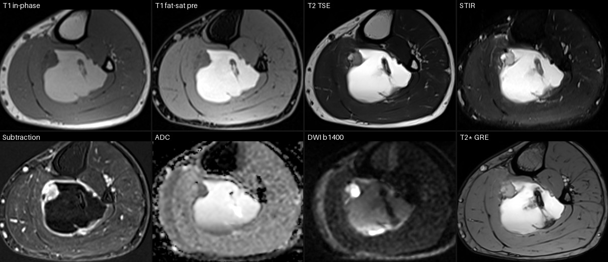
| Multiparametric MRI at 9.5 cm below joint | MRI 27 May 2026 · S24001, S27001, S19001, S17001, S1014001, S21001, S22001, S23001 · z -605 mm | Image-derived |
| Multiparametric MRI at 11.8 cm below joint | MRI 27 May 2026 · S24001, S27001, S19001, S17001, S1014001, S21001, S22001, S23001 · z -628 mm | Image-derived |
| Sagittal T1 TSE through the mass | MRI 27 May 2026 · S16001 · x 64.6 mm | Original |
| Sagittal post-contrast subtraction (scanner) | MRI 27 May 2026 · S1004001 · x 64.6 mm | Image-derived |
| Axial subtraction: transverse and AP diameters | MRI 27 May 2026 · S1014001 (post − pre, Dixon water) · z −608 | Image-derived |
| Sagittal subtraction: craniocaudal and AP diameters | MRI 27 May 2026 · S1014001 MPR · x 60 | Image-derived |
| Coronal subtraction: transverse and craniocaudal | MRI 27 May 2026 · S1014001 MPR · y 99 | Image-derived |
| Diffusion at the solid-component level | MRI 27 May 2026 · S22001 (b1400) | S21001 (ADC) · z −628 | Original |
| MRI 24 May vs MRI 27 May at matched levels below the knee joint line | Rows: 70, 90, 105, 120 mm below the tibial plateau. MRI-1 native slices (Study A S5; Study B S4/S8), MRI-2 S19001 / S1014001 / S27001 | Image-derived |
| Coronal STIR, both legs | MRI 24 May 2026 · Study A S4 Cor STIR B/L · im 16 | Original |
| Axial T2 at the widest level | MRI 24 May 2026 · Study A S5 Ax T2 · im 19 | Original |
| Subtraction (post − pre) | MRI 24 May 2026 · Study B S8 − S4 · im 18 | Image-derived |
| CT angiography, arterial phase, 7.8 cm below joint | CTA 27 May 2026 · S10 arterial 1 mm · im 990 · z 880 | Original |
| CT angiography, arterial phase, 9.6 cm below joint | CTA 27 May 2026 · S10 arterial 1 mm · im 1013 · z 862 | Original |
| CT angiography, arterial phase, 11.0 cm below joint | CTA 27 May 2026 · S10 arterial 1 mm · im 1030 · z 848 | Original |
| Arterial MIP (bone removed), frontal view | CTA 27 May 2026 · S10 arterial, bone masked with S4 (AI-derived MIP) · left knee to mid-calf | Image-derived |
| Arterial MIP (bone removed), lateral view | CTA 27 May 2026 · S10 arterial, bone masked with S4 (AI-derived MIP) · left knee to mid-calf | Image-derived |
| Subtraction angiography MIP, aorta to feet | CTA 27 May 2026 · S19 SUB-ANGIO · coronal MIP (AI-rendered) | Image-derived |
| Bone MIP, abdomen to feet (coverage) | CTA 27 May 2026 · S4 non-contrast · coronal MIP (AI-rendered) | Image-derived |
| HRCT chest 7 Sep 2026, coronal 9 mm MIPs | HRCT 7 Sep 2026 · S4 lung 1 mm · AI-rendered 9 mm coronal MIP at three depths | Image-derived |
| Low-dose CT chest 4 Jun 2026, coronal 9 mm MIPs | LDCT 4 Jun 2026 · S4 lung 1 mm · AI-rendered 9 mm coronal MIP | Image-derived |
| Upper and mid lungs, June vs September at matched levels | LDCT S4 vs HRCT S4/S10 · 9 mm axial MIP | Image-derived |
| Lower lungs, June vs September at matched levels | LDCT S4 vs HRCT S4/S10 · 9 mm axial MIP | Image-derived |
| Coronal STIR, both lower limbs: second-lesion survey | MRI 27 May 2026 · S6001 composite, slices 14/17/20/23 (raw and coil-normalised) · AI survey | Image-derived |
| Largest lymph node per station (short axis) | CTA 27 May 2026 · S15 late arterial · inguinal, external iliac, para-aortic | Image-derived |
| Femoral-head sclerotic foci (bone islands) | CT abdomen 4 Jun 2026 S4 and CTA 27 May 2026 S4 · same foci on both | Image-derived |
| Coronal STIR, both legs | Kathmandu MRI 24 May 2026 · Study A S4 Cor STIR B/L · im 16 | Original |
| Sagittal STIR, left leg | Kathmandu MRI 24 May 2026 · Study A S3 Sag STIR · im 10 | Original |
| Axial T2 at the widest level | Kathmandu MRI 24 May 2026 · Study A S5 Ax T2 · im 19 | Original |